

Apple Dental Centre | Ancaster, ON | appledentalcntr.ca
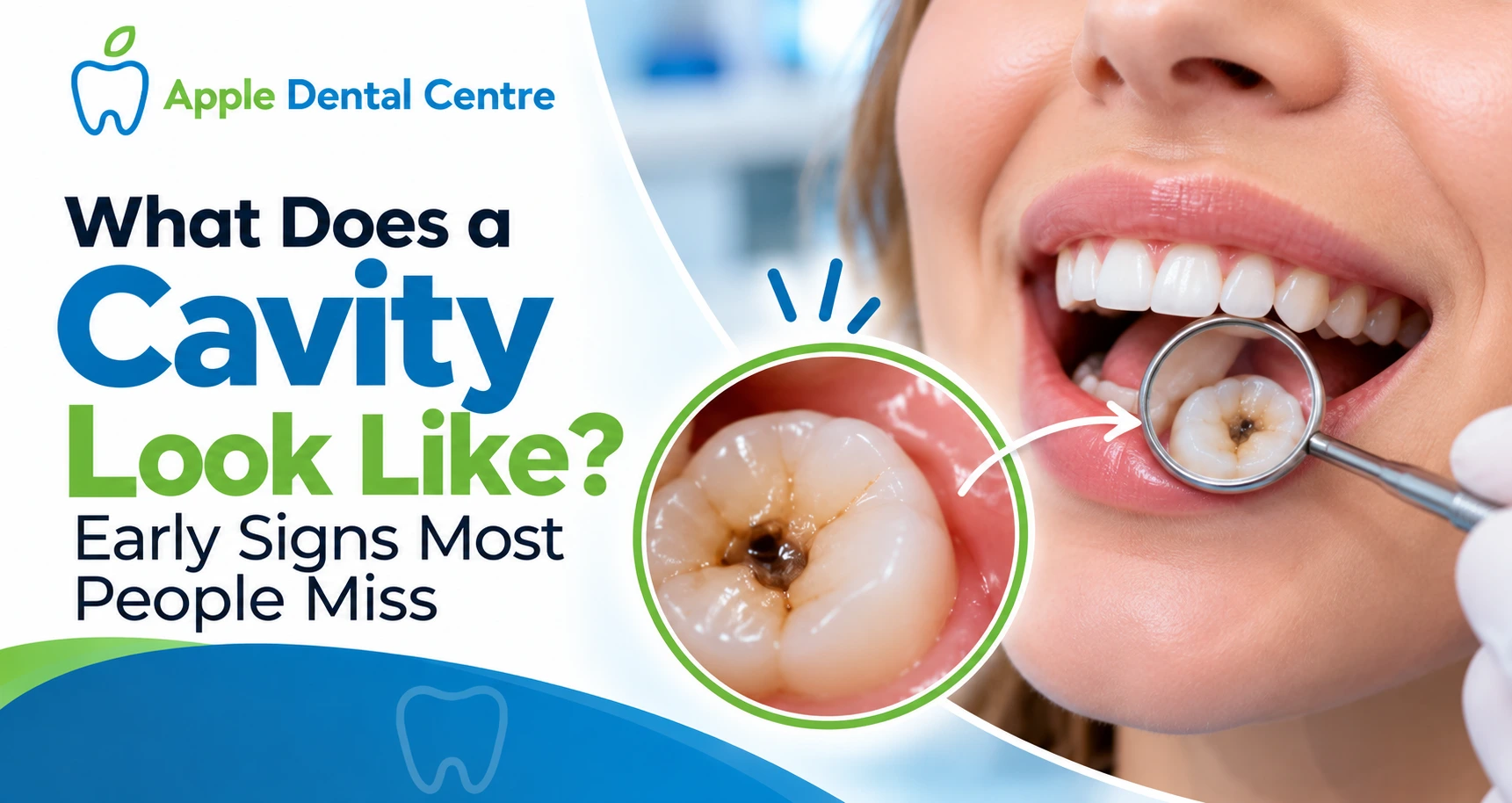
What Does a Cavity Look Like? Early Signs Most People Miss
Most people imagine a cavity as a dark, obvious hole in a tooth. In reality, the earliest — and most treatable — stages of tooth decay look nothing like that. They are subtle, easy to dismiss, and often invisible to the untrained eye entirely. At Apple Dental Centre in Ancaster, Dr. Subhi Alnahas, DDS, regularly sees patients who were completely unaware a cavity had been forming for months — sometimes because they were waiting for pain, and sometimes because they simply did not know what to look for.
This guide answers one of the most searched dental questions in detail: what does a cavity look like? We break it down by stage, by location, and by the subtle visual clues that most people miss until the damage has already deepened.
Understanding Cavities: What Is Actually Happening Inside Your Tooth
Before exploring what a cavity looks like visually, it helps to understand the biological process driving the damage. A cavity — clinically called dental caries — is not a single event. It is a progressive disease caused by acid-producing bacteria in the mouth.
When you eat sugars and refined carbohydrates, bacteria in your dental plaque feed on those sugars and release acid as a byproduct. That acid attacks and dissolves the minerals in your tooth enamel — the hard outer shell that protects the living layers beneath. This mineral loss is called demineralization, and it is the very first, still-reversible chapter of cavity formation.
Left unchecked, demineralization breaks through the enamel entirely and reaches the dentin — the softer, more porous layer beneath, where decay accelerates rapidly because dentin offers far less resistance than enamel. If decay is still ignored, it eventually reaches the pulp — the innermost chamber containing nerves and blood vessels — triggering significant pain and requiring a root canal treatment or even tooth extraction.
According to the National Institute of Dental and Craniofacial Research (NIDCR), dental caries is the most prevalent chronic disease worldwide — yet it is almost entirely preventable when caught early. The key is knowing what to look for before pain sets in.
Stage 1: What Does an Early Cavity Look Like?
The earliest visible sign of a cavity is not a hole and not a dark spot. It is a chalky white or opaque patch on the surface of the tooth enamel — commonly called a white spot lesion.
Healthy enamel has a smooth, slightly translucent quality. An early demineralization lesion disrupts that translucency, creating a flat, dull white area that does not catch the light the way the surrounding tooth does. These white spots most commonly appear:
- Along the gum line, where plaque accumulates most consistently
- On the smooth surfaces of front teeth, particularly in children and teenagers
- Beneath orthodontic brackets in patients who wear braces
This is the only stage where a cavity can be reversed. Fluoride — through toothpaste, professional fluoride treatments, and fluoridated water — can help remineralize the enamel before a true cavity forms. This is one of the most powerful arguments for regular professional dental cleanings and checkups in Ancaster — your dentist can identify and treat white spot lesions before they progress any further.
Stage 2: Yellow and Brown Discolouration — Decay Entering the Enamel
If a white spot lesion is not treated and remineralized, the area begins to absorb pigments from food, beverages, and bacteria — shifting in colour from white to yellow, then light brown. This colour change signals that the enamel surface has become porous and is actively breaking down.
At this stage, the cavity is still contained within the enamel layer. The tooth may not yet be painful, and there may be no physical hole visible. However, there is now a structural defect that will not reverse on its own. A small tooth-coloured dental filling at this stage is the ideal, minimally invasive intervention — the dentist removes only the decayed material and restores the tooth before the damage reaches the sensitive dentin below.
It is critical to distinguish between genuine staining from coffee, tea, or tobacco and active decay. Surface stains sit on top of the enamel and can be polished away during a professional cleaning. Decay spots are within the enamel structure and cannot be removed by cleaning alone. If you have noticed a new yellow or brown spot on a tooth, do not assume it is merely cosmetic — have it evaluated promptly by Dr. Alnahas and our Ancaster dental team.
Stage 3: Dark Brown and Black — Decay Into the Dentin
When decay breaks through the enamel and enters the dentin, the cavity darkens dramatically — appearing dark brown or black. The dentin layer is significantly softer than enamel, so once acid-producing bacteria reach it, the cavity expands more rapidly in all directions.
At this stage you may also notice:
- A visible pit or indentation on the tooth surface — especially on the chewing surfaces of molars
- Sensitivity to cold, heat, or sweet foods as decay approaches the nerve
- Pain when biting as the weakened tooth structure compresses under chewing pressure
- A rough or jagged texture when you run your tongue along the tooth
A cavity that has entered the dentin typically requires a larger dental filling or, if the damage is extensive, a dental crown to restore the tooth’s structural integrity. The earlier you act within this stage, the better the outcome — waiting further risks pulp involvement.
What Does a Cavity Between Teeth Look Like?
Interproximal cavities — cavities that form between two teeth — are among the most commonly missed because they are almost entirely hidden from view. They develop in the contact zone where two teeth meet, an area that is impossible to clean with a toothbrush alone and where plaque accumulates if flossing is infrequent.
Visually, you will rarely see anything at all in the early stages of an interproximal cavity. In more advanced cases, you may notice:
- A subtle grey shadow visible just below the contact point when looking carefully in bright light
- A small dark triangle at the gum line between two teeth
- Floss tearing, shredding, or catching consistently in the same location
- Mild cold or sweet sensitivity in the area
The definitive way to detect interproximal cavities is with bitewing X-rays — the targeted dental radiographs that show the contact zones between teeth in cross-section. At Apple Dental Centre, we use modern digital dental radiographs that reveal interproximal decay at its earliest detectable stage, with minimal radiation exposure.
What Does a Cavity on a Front Tooth Look Like?
Cavities on front teeth — the incisors and canines — are particularly distressing because they are visible when you smile. Fortunately, they are also easier to spot early because of their accessibility. On front teeth, look for:
- White or chalky patches on the smooth front surface
- Brown or dark lines along the edge where two front teeth touch each other
- A translucent or glassy appearance along the biting edge
- A small chip or notch on the biting edge where weakened enamel has fractured away
Front tooth cavities often develop from prolonged exposure to acidic beverages, poor brushing technique, or dry mouth. A tooth-coloured composite dental filling can restore a front tooth discreetly, matching your natural tooth shade.
When Visual Signs Are Absent: Physical Warning Clues
Many cavities — particularly early-stage and interproximal ones — produce no visible signs at all. In these cases, pay attention to:
- A brief sharp zing when eating something cold, hot, or sweet
- Prolonged sensitivity to temperature changes
- A mild ache in a specific tooth without an obvious cause
- Food consistently getting stuck in the same spot between two teeth
- A new roughness or sharpness on a tooth surface
For a comprehensive overview of the sensory warning signs of decay, read our detailed guide: How to Know If You Have a Cavity: 10 Warning Signs to Watch.
If you experience any of these symptoms alongside the visual signs described in this article, do not wait. If pain has become severe or you notice facial swelling, contact our Ancaster emergency dental care team.
How Dentists Confirm What They See: The Diagnostic Process
Even when a cavity seems obvious visually, professional confirmation is essential before treatment. At Apple Dental Centre, Dr. Alnahas uses a multi-tool diagnostic process to assess suspected areas of decay:
- Visual and tactile examination: Using magnification and dental instruments to assess areas of suspected decay.
- Digital bitewing X-rays: Our dental radiographs can reveal interproximal cavities and decay that may not be visible during a visual exam.
- Transillumination: Directing a focused beam of light through the tooth to reveal areas where decay may have disrupted the tooth’s mineral structure.
The combination of these methods helps the dental team develop an appropriate treatment plan while preserving as much healthy tooth structure as possible.
What Happens If You Ignore a Cavity?
Untreated decay can progress from enamel to dentin and, eventually, toward the pulp. Treatment needs can become more extensive as decay progresses.
- White spot stage: Fluoride treatment and preventive care may help remineralize early enamel changes.
- Enamel cavity: A small dental filling may be required.
- Dentin cavity: A larger filling or dental crown may be needed depending on the extent of damage.
- Pulp involvement: Root canal treatment may be required.
- Severe infection or fracture: Tooth extraction may be necessary, followed by replacement options such as a dental implant or bridge.
About Apple Dental Centre — Expert Cavity Care in Ancaster
At Apple Dental Centre, we believe that an informed patient is an empowered one. Dr. Subhi Alnahas, DDS, brings over 14 years of clinical experience in restorative dentistry and cavity treatment to every appointment. Our clinic is located at 73 Wilson St W, Unit 20, Ancaster, ON L9G 1N1, and we serve patients across Ancaster, Hamilton, Dundas, Stoney Creek, and Waterdown.
We accept the Canadian Dental Care Plan (CDCP) — meaning eligible patients may have their dental fillings and preventive care fully or partially covered. Learn more about CDCP coverage at our Ancaster clinic.
Professional dental cleanings are available from just $99 at Apple Dental Centre.
Meet Dr. Alnahas and our team | View all dental services | Book your appointment online today
Frequently Asked Questions About What a Cavity Looks Like
Expert answers to the questions Ancaster patients ask most about cavity appearance, stages, and treatment.
Do not wait for a dark hole or severe pain to tell you something is wrong. If you notice a change in your tooth, experience sensitivity, or are concerned about possible decay, arrange a professional dental examination.
Call us at (289) 204-9090 or book your appointment online.
📞 Call us at (289) 204-9090
💻 Book your appointment online today.
Apple Dental Centre | 73 Wilson St W, Unit 20, Ancaster, ON L9G 1N1 | (289) 204-9090 | appledentalcntr.ca